Brain tumor biopsy
The content of the article
A brain biopsy is a diagnostic procedure that involves removing a small sample of tissue ( biopsy ) for further study. The data obtained allows us to establish the necessary steps for subsequent treatment. Doctors decide on the methodology - whether surgery, chemotherapy, radiation therapy , etc.
Taking material helps to determine the degree of malignancy of the tumor. In addition, this process is often prescribed to confirm Alzheimer's or Creutzfeldt -Jakob disease. Using a biopsy, healthcare professionals also diagnose multiple sclerosis and inflammatory processes in the brain.
In standard cases, this type of diagnostic study is carried out at the last stage and in conditions of insufficient information collected by other methods.
A biopsy becomes unnecessary: when the last stage of cancer development has already started, there is an acute period of inflammatory processes, as well as in case of blood clotting problems.
Experienced neurosurgeons use the following types of brain biopsies:
- Open.
- Stereotactic.
- puncture.
Open brain biopsy
The peculiarity of this type of biopsy is that the collection of material ( biopsy ) from brain tissue occurs directly during the operation. Among other types of diagnostics, it is the most dangerous, since neurosurgeons have to perform many manipulations. But at the same time, this is a fairly common method. So, tissue collection occurs as follows:
- A hole is made in the skull.
- Part of the bone is removed, leaving a window for trephination.
- Through it, the formation is removed.
- The open area is covered with bone or a special surgical plate.
- A biopsy is taken from the removed tumor.
If a person has pronounced cerebral edema, the defect remains open and is covered with a skin flap. This decision is made by doctors depending on the severity of the disease and the danger of possible infection.
Stereotactic biopsy
Stereotactic biopsy involves a minimally invasive procedure. The process of collecting brain tissue consists of the following steps:
- A person is put on a stereotactic frame, which allows him to hold his head in the desired position.
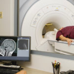
- Surgical intervention is accompanied by CT or MRI . Using a modern device, the doctor can enter the results directly into a special program.
- A cut of up to 1 cm in size is made in a pre-prepared locus on the skull, after which is drilled .
- A puncture needle, which is equipped with a microcamera , is inserted into the resulting hole . The recording camera transmits the image online . With its help, the doctor can examine in detail the formation and the required location for the puncture.
- A biopsy sample is taken and the hole is sutured.
Needle biopsy
A puncture biopsy of the brain involves making a small hole in the skull and using a needle to remove a small amount of tumor or brain tissue. It is the least dangerous type of surgery. The process is as follows:
- At the site of the suspected formation, a skin incision is made on the head.
- Use a drill to make a corresponding hole.
- Penetrate into the cranial cavity with a puncture needle.
- The biopsy is taken from a specific point of the tumor.
Among the three types of brain biopsy, it is worth highlighting: stereotactic and puncture , since they are the most modern and least traumatic methods. After such operations, a person can go home on the first day, and on the 7th day it is possible to do his usual activities.
Please rate the article:


