Calcified meningioma: diagnosis and method of its treatment.
The content of the article
In medical practice, meningioma is often found on the surface of the brain (outside the brain), but there are cases of tumor formation in other parts of the brain. Meningioma develops quite slowly and rarely develops from benign to malignant.
Causes of tumor appearance.
Scientists are in a difficult position, because it is not yet possible to establish the cause of meningioma. However, it is possible to identify the factors that provoke its development. These factors are:
- Exposure to ionizing radiation;
- Deviation in the development of chromosome 22;
- Mature age (40 years and older);
- Brain injuries.
Classification differences.
Classification of meningioma:
- Angiomatous;
- Chordoid;
- Secretory;
- Psammomatous;
- Meningotheliomatous;
- Petroclival;
- Hyperostotic olfactory;
- Calcified;
- Transitional.
It is possible to classify meningioma only after undergoing comprehensive diagnostics. There are also three categories of meningioma:
- Benign (typical) - a superficial neoplasm that slowly grows into the brain tissue, causing its compression.
- Atypical ( semi-benign ) is a neoplasm that tends to grow into brain tissue and is characterized by mitotic growth activity.
- Malignant ( anaplastic ) - the most dangerous type of tumor, penetrates into the brain tissue. Leads to the formation of metastases.
Manifestation of meningioma .
At the first stage of development, there may be a complete absence of symptoms. Their manifestation is possible only after the meningioma has acquired a certain size.
The symptoms are:
- Cephalgia;
- Increased blood pressure;
- Nausea;
- Memory losses;
- Weakness, loss of balance;
- Deviation in the psyche;
- Impaired hearing, vision and smell.
Also, the symptoms depend directly on the location of the tumor:
- Superficial formation (Headaches that worsen after waking up and before bedtime, cramps).
- Education in the frontal lobe (Changes in a person’s mental state, impaired vision and smell).
- Neoplasm of the temporal region (deterioration of hearing and vision, confused speech, weakness, seizures).
- Tumor in the parasagittal region (Affects hearing and coordination).
- Cerebellar neoplasm (hearing impairment, respiratory depression).
- Neoplasm in the area of the sella turcica (complete loss of vision).
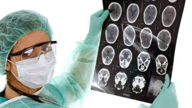
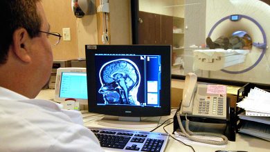
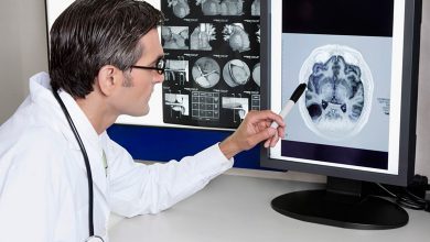
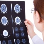
Diagnosis of neoplasm.
In the early stages, diagnosis of swelling is difficult. In many cases, the symptoms are similar to age-related diseases. Diagnosing meningioma is possible only after undergoing a comprehensive and comprehensive medical examination. The examination must be carried out under the supervision of the following specialists: a neurosurgeon; neurologists; otolaryngologist and ophthalmologist. When symptoms are identified, the following procedures are performed to confirm the diagnosis: computed tomography and magnetic resonance imaging; blood test and biopsy.
Tumor treatment.
Treatment of the tumor is possible only after a complete diagnosis. Considering the location, size and degree of malignancy.
The most common methods of treating meningioma:
- Medical support is a conservative method of treatment. This method is only possible if the tumor is not malignant and does not affect the patient’s well-being.
- Surgical method ( meningiolysis ) – removal of non-cancerous tumors through surgical resection. This method is used to remove large tumors.
- Radiation therapy is a technique that allows you to remove a malignant tumor that has many locations. Therapy is carried out repeatedly. This method has a large number of side effects (radiation dermatitis, alopecia, deterioration of the patient’s condition). It is used only if the tumor cannot be removed by surgery.
- Gamma knife removal – used for small tumors. The tumor is removed using a concentrated stream of ionizing radiation. This method does not affect healthy cells and is minimally invasive.
Please rate the article:
 (2 ratings, average: 5,00 out of 5)
-
 Read also: Is disability given after removal of brain meningioma
? Open article

The list was formed on the basis of the lack of convincing data on the effectiveness of drugs for the stated indications, as required by evidence-based medicine, as well as the absence of authoritative sources and recommendations.