Glioblastoma of the brain
The content of the article
The most common primary brain tumor is glioblastoma. It is highly aggressive and malignant in nature and originates from neuroglia. Despite the lack of metastasis, this brain tumor still remains the leader in terms of the most dangerous tumors. Even if glioblastoma of the brain is treated, the prognosis after surgery for most patients remains unfavorable.
Brain tumor stage 4
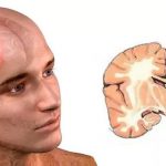
Stage 4 glioblastoma of the brain, photos of patients of which can be found on the Internet, is formed as a result of the active proliferation of astrocytes. This brain tumor is characterized by a chaotic accumulation of tumor cells with polymorphic nuclei. Glioblastoma is extremely life-threatening for the patient precisely because of the active growth of the tumor, as well as the lack of a clear boundary between the affected and healthy tissues. The rapid proliferation of tumor cells leads to the fact that they begin to compress the brain, and then grow into the tissue, thereby destroying it.
Glioblastoma: causes of occurrence
To date, experts have not yet established the exact reasons that result in uncontrolled proliferation of glial cells, and as a result, the development of glioblastoma. There are only guesses on this score: it is believed that ostrocytes are negatively affected by electromagnetic radiation, which affects a person as a result of active use of a mobile phone, but this theory could not be scientifically confirmed.
At-risk groups
- Men over 40. Statistics show that men from 40 to 60 years old suffer from this disease much more often than at any other age. In the second half of humanity, glioblastoma is much less common;
- Presence of astrocytomas . More than 10% of diagnosed fibrillary and pilocytic astrocytomas, which are not very malignant, subsequently degenerate into glioblastoma;
- Neurofibromatosis. Most often, glioblastoma occurs in patients who suffer from neurofibromatosis. The neoplasm occurs as a result of disorders at the genetic level;
- Ionizing radiation and exposure to polyvinyl chloride. Other chemicals also have a very negative effect on clial cells, which leads to malignant formation;
- Viral diseases;
- Hereditary predisposition;
- Penetration of infected cells into the cerebral cortex through the circulatory system.
Kinds
These neoplasms are usually classified according to histological criteria. As a result of diagnosis, one of the following types of glioblastoma is determined:
- Giant cell. A sample of this type of tumor will be distinguished by a large number of huge cells, the nuclei of which will be clearly visible under a microscope and will be polymorphic;
- Multiform. In this case, the sample will differ not only in the large number of malignant cells, which will be localized in a chaotic manner and differ in different shapes, but also by the presence of proliferation of blood vessels. Under a microscope, a specialist will clearly see focal necrotic lesions. This subtype belongs to grade 4 and is the most dangerous with a malignant nature;
- Gliosarcoma. It is distinguished by bidermality, which means a mixture of glial and mesenchymal tumors;
- Polymorphocellular. This form is diagnosed quite often. Thanks to cytological methods, it is possible to quickly detect tumors of various shapes and sizes. Compared to other structures, the cytoplasm of this area takes up little space and is weakly stained during examination. Cell nuclei differ in structure. There are mainly oval, round, bean-shaped and irregular shapes. This type of tumor in some cases can be classified as giant cell glioblastoma;
- Isomorphic cellular. I diagnose this form quite rarely. Basically, the tumor cells are uniform, but there are still slight differences in the shape and size of the cells;
- Glioblastoma of the brain stem. When this type of tumor is diagnosed, the prognosis is unfavorable. Such a pathology cannot be subject to surgical treatment, since at the site of localization there are very important structures that are responsible for important functions of the body. This trunk connects the brain and spinal cord. It contains the cranial nerves, as well as the vasomotor and respiratory centers. That is why, when diagnosing this type of tumor, special attention is paid to the patient’s circulatory and respiratory disorders. The tumor can begin to develop from the brain stem itself or from another part of the brain. The development of infected cells occurs at a rapid level, and the cells in it are atypical.
Symptoms
It is worth noting that when diagnosing a disease such as glioblastoma of the brain, the symptoms will be very extensive. Already at an early stage of development of the tumor, especially if it is localized in the centers of the brain that are responsible for speech and movement, severe problems with speech, fainting, and uncontrollable problems with movement may be observed.
In some situations, when part of the brain has already been affected by glioblastoma, symptoms of the disease may be completely absent. In most cases, in the presence of a neoplasm such as glioblastoma, symptoms will appear in the early stages of the disease. Symptoms will directly depend on the location of the tumor, as well as its size.
There are several possible manifestations of this disease:
- Changes in character and frequent mood swings;
- Memory impairment;
- Violation of visual and auditory processes;
- Decreased attentiveness and concentration;
- Impaired sensitivity;
- Motor impairment;
- Impaired functioning of the limbs. If the tumor affects the right hemisphere of the brain, the left leg and arm will be affected. If the left hemisphere is damaged, the right arm and leg will suffer accordingly. Most often, patients complain of weakness in the limbs, but partial paralysis can also be diagnosed;
- Violation of thought processes;
- Decreased learning ability;
- Headache. The main symptom is severe periodic headache , but as the tumor grows, the headache can become constant;
- Decreased appetite. As a result of a brain tumor, the patient may lose a lot of weight, but most often this is not given much importance;
- Dizziness;
- Epileptic seizures, which do not occur so often and depend on the location of the formation;
- Increased sleepiness.
Diagnostics
To establish a diagnosis of a brain tumor, the following is required:
- Neurological examination of the patient;
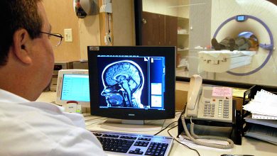
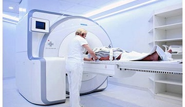
- MRI and CT of the brain. What it is? This is magnetic resonance or computed tomography , which allows the specialist to obtain the most complete picture of the patient’s illness;
- Antiography of cerebral vessels. This procedure is prescribed only if necessary and if surgical intervention by a specialist is required. This method allows you to study blood circulation in the tumor tissue;
- PET-CT. This is a fairly new diagnostic method, which has already proven its effectiveness in detecting and diagnosing brain tumors.
Treatment
All treatment methods for diagnosing glioblastoma are aimed at either completely removing the tumor or partially reducing its activity, which is possible due to the absence of metastases.
The entire course can be divided into three stages:
- Surgical intervention;
- Radiation and chemotherapy ;
- Maintenance chemotherapy.
Surgical intervention
When diagnosing a disease such as glioblastoma of the brain, life expectancy depends most of all on the possibility of complete removal of the tumor by surgery. The highest effectiveness of complex effects on the tumor is achieved only after neurosurgical intervention.
Until recently, even highly qualified specialists with extensive experience could not completely remove the tumor due to its growth into the brain tissue, as well as entanglement of the patients’ vital centers with the affected cells. As a result, it was possible to remove only particles of infected cells, which led to the completion of the operation without achieving the necessary results.
The patient is prepared for surgery 4 hours before it begins. He is given a special solution to drink, which is based on this same 5-aminolevulinic acid. After this, the process of protoporphyrin accumulation begins in tumor cells. This is what allows the contours of dead cells to glow.
Radiation therapy
After surgery, the patient is sent for radiation therapy. Surgical intervention cannot bring 100% results, and therefore malignant cells can continue to multiply in the lesion. In a small proportion, radiation therapy can replace surgery. This occurs in cases where the disease is inoperable . The procedure is quite lengthy, so to achieve the desired result, the patient is prescribed 6-week therapy, 5 times a week. The total number of rays during the entire procedure is about 60 gray. This dose is considered the most optimal, as it has an effective effect on malignant cells and does not cause negative consequences for the patient. Throughout the course of treatment, the patient must take the special antitumor drug Temodal daily.
The use of this therapy also has its side effects, which may manifest themselves to a lesser or greater extent. The patient may feel fatigue, nausea, and suffer from excessive hair loss. Brain tissue may swell greatly, leading to intermittent or constant pain. Under the influence of radiation, necrotic processes can form.
Chemotherapy
The patient also needs supportive therapy. In this case, specialists again prescribe the already known drug Temodal. The antitumor drug Temodal is re-prescribed to the patient 4 or more weeks after the end of the second stage of treatment, that is, radiation therapy. This stage is also quite lengthy, since the patient must take the drug in courses of five days. As a result, 6 such courses are prescribed with a break of 23 days.
Targeted therapy
This may include taking a new drug called Avastin. It has an effective destructive effect on the tumor. This drug destroys tumor vessels, and glial cells lose their ability to reproduce.
At the moment, this drug is not used for the treatment of primary tumors, since it has not proven its 100% ability to destroy affected glial cells. But its effectiveness in treating relapses has been scientifically proven.
Forecast and consequences
Most of all, patients and close relatives are concerned about the following questions: what is glioblastoma of the brain, how long do they live with it after surgery, and what is the chance of a full recovery? Unfortunately, due to the malignant nature of this neoplasm, the prognosis is extremely unfavorable even for those patients who have completed the entire course of treatment. The time at which the tumor is diagnosed has a very important impact on the prognosis.
Statistics show that only 10% of all patients diagnosed with this disease have a chance to live up to two years after diagnosis.
The most dangerous form of the disease is grade 4 glioblastoma of the brain, the prognosis of which is very unfavorable - if treatment is successful, the patient is not given a chance to live even 40 weeks. Today there have been practically no cases where a patient was able to live more than five years after diagnosis. The most unpleasant answer awaits those who are interested in the question: how long do people live with stage 4 glioblastoma?
The very low survival rate is associated with the following consequences after treatment of this disease:
- Even if the primary tumor is successfully treated, about 80% of patients experience recurrence;
- Often the tumor is localized in particularly important centers of the brain and begins to actively affect healthy tissue, which makes it impossible to remove the tumor completely. As a result, malignant cells affect the centers that are responsible for blood circulation and respiration;
- If it is impossible to completely remove the tumor, malignant cells lead to neurological disorders or complete paralysis of the patient's body.
The development of oncology and neurosurgery does not stand still. We cannot exclude the possibility that in the near future, with the help of modern developments in the field of medicine, it will be possible to successfully cure patients who suffer from a similar disease, especially considering the fact that until recently, complete removal of glioblastoma was impossible.
Please rate the article:

 (6 ratings, average: 4,17 out of 5)


