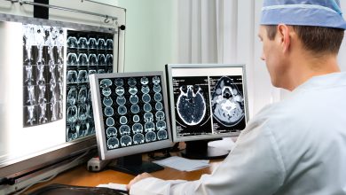
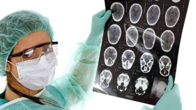
CT scan of brain tumor
The content of the article
The most complex and most important organ of the human body is the brain. In view of this, in case of suspicion of the presence of diseases of this organ, doctors do everything possible to quickly and as accurately as possible make a diagnosis. For quite a long time, the most reliable way to diagnose a brain tumor has been a CT scan.
CT scan of brain tumor
This is a diagnostic technique based on the effect of X-ray radiation on the brain structures. X-rays produced in the X-ray tube of a tomograph affect the area of the patient’s body being studied from different angles. The sensors contained in this equipment receive information about layer-by-layer scanning, which is subject to immediate processing on a PC and make it possible to “build” an overall picture of the development of the tumor. In this case, the specialist is presented with a detailed image of the brain, allowing an accurate diagnosis to be made with great confidence.
A distinctive feature of the X-ray apparatus is computer diagnostics of brain tumors. It is more informative and allows you to study the organ in detail.
At the same time, computer diagnostics of brain tumors, unlike MRI , is cheaper and has no contraindications for use: the presence of metal prostheses, implants, or a pacemaker in patients does not prevent CT from being performed. One of the ways to study tumor tumors is puncture of a brain tumor. It is used to detect pustular formations.
During the procedure, the development of complications cannot be ruled out:
- the brain may become infected; vascular damage cannot be ruled out;
- pus may end up in the ventricles of the brain. To eliminate all kinds of complications during the procedure, careful adherence to the rules is necessary:
- disinfection and treatment of the dura mater with peroxide, followed by iodine;
- to exclude cases of vascular damage, a special needle with a blunt tip is used;
- the procedure is carried out at a certain depth (no more than 40 mm), this will exclude cases of pus penetrating into the lateral ventricles of the medulla. Several needles are prepared for the procedure in case one of the instruments used becomes clogged with brain tissue. The needle must have certain width parameters. Not every instrument can remove pus.
The most suitable tool is a special needle equipped with a mandrel.
What diagnostics are performed for a tumor?
The basis of CT work is the effect of X-ray radiation on tissues and organs. When working with a CT scan, a specialist analyzes the three-dimensional image of an organ obtained by the equipment. It's all about the structure of the device itself: the source of X-ray radiation is a ring-shaped circuit, inside of which there is a couch (table) for the patient. Thus, the device makes it possible to take a number of X-ray photos of organs that have different angles and are located in different ways.
Image processing is carried out using a computer, and a three-dimensional model of the organ is recreated based on the obtained material. The most important aspect of the study is that the specialist has the opportunity to view “slices” of the organ, depending on the options used in the device; the slice thickness parameters can be up to 1 mm, which significantly increases the accuracy of the diagnostic procedure. MRI is based on the same principle. The studies differ in the nature of the waves generated: in the case of MRI, they are electromagnetic.
Under the influence of wave healing of this type on different organs and systems, a varied response is obtained, recorded by acceptable devices of the apparatus. And after, as after CT, the signals are processed and converted into an image. There are differences between CT and MRI. First of all, they consist in what pathologies are detected using one or another diagnostic method, as well as in the rays produced by these devices. Whether a CT scan will show a brain tumor is an important question that can be answered by a professional or special literature. To resolve this issue, you need to understand in what situations this procedure is prescribed.
- hemorrhages and strokes;
- fractures of the base of the skull;
- bleeding;
- trauma and hematomas;
- neoplasms and cysts;
- encephalitis and abscesses;
- aneurysms;
- abnormal development;
- presence of foreign bodies.
In addition to the main version of CT examination, it is possible to talk about its variety - CT angiography of the brain.
A similar study is prescribed for:
- diagnosing cerebrovascular disease:
- in case of interruptions in blood circulation to brain structures;
- presence of neoplasms.
CT angiography is classified depending on the type of vessels studied:
- venography;
- arteriography.
Preparing for CT
The patient is referred to the CT scan by a doctor. There can be many reasons for conducting research:
- getting head injuries;
- chronic migraines;
- loss and confusion of consciousness;
- hearing and vision impairment;
- frequent dizziness, tinnitus;
- increased level of intracranial pressure;
- for the purpose of monitoring the treatment of pathologies.
The procedure is contraindicated for pregnant women and children. Assigned to these categories of persons in cases where the life or death of the patient is at stake. The procedure is not advisable for nursing mothers, but it is quite possible if there are indications for it. If the diagnostic procedure is carried out using a contrast agent, breastfeeding should be avoided for 24 hours. Persons whose body weight is more than 150 kg are not allowed to undergo CT examination due to the peculiarities of this equipment.
You can take this test at any time of the day. There are contraindications to the procedure when using contrast - before the procedure, the patient takes a blood test for urea and creatine levels, sometimes it is necessary to limit food intake 4 hours before the test.
When conducting an examination in the office, the patient changes into a disposable shirt. It is necessary to remove jewelry and lie down on the mobile tomograph table. The patient's head is fixed and the table is placed in the scanner. After the device is started, noise and clicking noises are heard. The patient remains in a static position until the study is completed; if necessary, he can consult a doctor.
Pituitary tumors, which is better: MRI or CT?
This question concerns most patients who develop intracranial sarcomas. The process of treating these tumors is extremely complex due to the fact that the so-called “sella turcica” (pituitary gland) is one of the most important endocrine glands. This small organ is located at the base of the brain; its role in human life is significant, because the pituitary gland is the central endocrine gland.
When preparing for an MRI of the pituitary gland, eating before the study is not so important. If the procedure is carried out using a contrast agent, it makes sense to abstain from food for some time before the study, which will eliminate the occurrence of side effects - nausea, vomiting. The specialist must know in advance information about the presence of allergic reactions to contrast and asthma in the patient.
Preparing for an MRI
During this research procedure, there is a high-frequency magnetic field in a separate room; therefore, the patient, when visiting the MRI room, must comply with the following rules:
- jewelry, payment cards, hearing aids may be faulty;
- small metal products: pins, hairpins, zippers can worsen the quality of the procedure;
- removable dentures with metal elements are recommended to be removed;
- It is not recommended to enter the office with pens, pocket knives and glasses;
- The piercing is removed in advance.
The procedure has a number of advantages:
- MRI does not use ionizing radiation;
- With MRI, the image of the pituitary gland is clearer and more detailed than images obtained using other methods of visualizing the organ;
- MRI is applicable in diagnosing pathological conditions at an early stage of development, including neoplasms.
- When performing an MRI, the doctor receives information about the structural structure of the organ, and sometimes about the functions performed by the pituitary gland.
If there are certain mental disorders of the patient, in particular claustrophobia, it is possible to conduct research using open-type equipment. But with innovations in the production of open-type apparatus using high-frequency magnetic waves, the quality of research has increased.
Please rate the article:
 (2 ratings, average: 5,00 out of 5)
-
 Read also: Dexamethasone for brain tumors
Open article

The list was formed on the basis of the lack of convincing data on the effectiveness of drugs for the stated indications, as required by evidence-based medicine, as well as the absence of authoritative sources and recommendations.