Oligodendroglioma
The content of the article
Brain tumors are classified as serious diseases, and oligodendroglioma is one of them.
This tumor arises from those fat cells that make up the membrane of nerve cells.
Doctors classify these types of brain tumors as primary glial tumors, and they are subdivided based on the degree of malignancy. According to the WHO classification there are II and III degrees. Their difference is that each degree has its own course - slow or progressive, and patients live a certain number of years after the first symptoms of the disease began to appear. The disease is considered less severe when considering the statistics of brain parenchyma tumors, due to the fact that such a tumor is less aggressive in terms of biological behavior and is treatable when chemotherapy is used.
But still, this type of brain tumor is most often detected in middle age, after the age of 40, and men most often suffer from it. Brain oligodendroglioma arises from oligodendrocytes. It can be detected in the white matter of the cerebral hemispheres, but not immediately, since it grows rather slowly, however, it can reach large sizes.
Its favorite location is along the walls of the ventricles. As the tumor grows, it begins to enter their cavity, then penetrates the cerebral cortex.
Oligodendroglioma
Quite rarely, but still such a tumor can occur:
- in the brain stem;
- in the optic nerve;
- in the cerebellum.
An experienced doctor can identify a tumor by criteria such as clearly defined borders and a pink, pale color. Cysts may appear inside . They grow very slowly, but since cysts can grow to large sizes, they significantly complicate treatment.
Oligodendroglioma has its own specific characteristics, for example, expansive-infiltrative growth. But before a diagnosis is made, it may take up to 5 years, since the tumor has a long symptomatic course, during which loss of heterozygosity of chromosome 19q occurs.
Patients are concerned about whether they will recover in the future. The prognosis depends on the location of the tumor and how the brain structures in the immediate vicinity behave. On average, having decided on a diagnosis, doctors believe that complex treatment prolongs the life of patients by up to 10 years if the oligodendroglioma is highly differentiated, and only up to four years if the tumor is classified as anaplastic.
Symptoms
There are a number of symptoms by which a doctor can diagnose oligodendroglioma in absentia. These primarily include:
- constant headaches that occur in attacks;
- numbness of the limbs;
- seizures, which occur in almost 90 percent;
- movement coordination disorder;
- over time, behavioral changes, disturbances in reactions;
- seizures similar to epileptic ones;
- hemiparesis.
It is seizures that are considered nonspecific symptoms. Some patients have generalized seizures, others have partial seizures, and there is also a mixed type. The younger the patient, the more worried he is about constant convulsions, therefore, by going to the hospital with such a problem, the diagnosis can be detected much earlier - after diagnosis. Older people most often experience neurological symptoms.
Causes of the disease
Unfortunately, the nature of the occurrence of many tumors has not yet been fully studied. The reasons for the mutations that occur have not been determined.
At this stage, data have been collected by which we can judge what exactly provokes the appearance of oligodendroglioma in the brain. Those most often at risk are:
- men;
- people whose age category is over 40 years;
- patients whose relatives have experienced the same disease. Experiments were conducted that proved that there is a genetic predisposition to the disease, and mutations at the cellular level can appear in a person with such pathologies in genes;
- patients who have been exposed to radiation therapy;
- working professions if you have to constantly work with toxic and chemical substances;
- infection with viruses that can have increased oncogenicity.
Classification
Doctors classified oligodendroglioma tumors, dividing them into 2 classes. This:
- low-grade (slow and limited growth);
- anaplastic oligodendroglioma. A dangerous malignant formation that tends to rapidly grow malignant tissue.
The clinical picture can be observed in patients quite similar. For those who are susceptible to a slow-growing type of pathology, the symptoms are erased or almost not observed.
Further development of the disease is characterized by the occurrence of headache. The disease will also affect emotional disturbances.
Pathology can occur in parts of the brain such as the temporal lobes. In this case, memory deteriorates significantly, speech becomes slurred, and coordination of movements is impaired. If a tumor develops in the area of the frontal lobes, the person experiences constant weakness, and half of the body begins to go numb.
General malaise is accompanied by increased intracranial pressure, resulting in pain, nausea, and often blurred vision. In the brain, oligoastrocytoma can occur, which is of the glial type, and its development is facilitated by small astrocyte cells. Their usual task is to maintain brain cells in working order, and this type of cell is classified as glial.
The consequences of the disease cause difficulties for the patient over time. If the tumor arose in the right hemisphere, over time there is a possibility of paresis in the left limbs. If the left hemisphere is damaged, the right side will feel the consequences.
Developing in the cerebellum, the tumor will impair coordination of movements, and if the pathology develops in the occipital lobe, the person’s vision decreases and hallucinations may occur. When the tumor is located in the frontal lobe, a change in character is observed, the patient becomes hot-tempered and unbalanced.
Four degrees of severity
Brain oligoastrocytoma is divided into four groups depending on the degree of danger to human health:
- policytic. It has I degree of malignancy, therefore it is not particularly dangerous and is classified as benign. It develops slowly and has clear contours. Diagnosed in children in the cerebellum or brain stem. An experienced doctor can remove it. There is no further impact on health;
- fibrillar. She has stage II malignancy. It is also benign and also grows slowly. But it is more difficult to remove it, since the boundaries are not so clearly marked. Most often it affects young people aged 20 to 30 years;
- anaplastic. It belongs to the III degree and is already a malignant formation. Its development occurs quickly, and it is very difficult to respond to surgical treatment due to blurred contours. The severity of this disease is experienced in most cases by middle-aged men;
- glioblastoma. This is the most dangerous type of disease. It has dangerous properties - it grows into brain tissue, resulting in brain necrosis. It is most often diagnosed in the age group from 40 to 70 years.
Malignant oligodendrogliomas, which have stages III and IV, behave in the same way as astrocytomas .
Preliminary studies
They are needed so that once a brain oligodendroglioma has been identified, complete recovery is successful. Patients will have to go through several stages of the study.
First, when visiting a medical institution, a primary examination takes place. The doctor is obliged to find out whether the back surface of the eyeballs and nerve reflexes are in order. Finds out what pathologies are occurring with the patient and what worries him most.
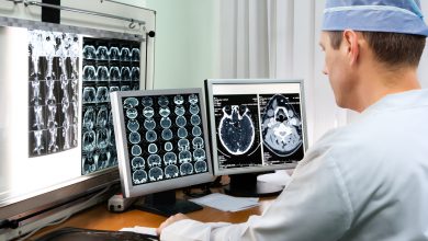
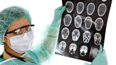
It is necessary to undergo examination using computed tomography. At its core, this is an x-ray scan of the area of the brain that has been affected by the tumor. During the examination, the specialist examines a three-dimensional image depicting brain tissue. To ensure clearer image quality, the patient takes a special contrast agent before the procedure, which is harmless to health. The examination takes about 30 minutes and does not cause any pain.
Magnetic resonance imaging is used to make a correct diagnosis. The procedure is based on the effect of magnetism. At the first stage, the subject is injected with a dye, with the help of which a detailed image of the diseased area of the brain is obtained clearly.
For histological analysis (biopsy), a small piece is taken from the affected area. This research method is performed surgically, but the feasibility of this method is not always justified; it all depends on the individual case.
Tumor treatment
In order for an identified brain oligoastrocytoma to have a positive prognosis, it must be treated urgently. The choice of the correct therapy depends on the immediate location, size of the tumor and its specific nature.
The task of preparing a comprehensive treatment is usually assigned to doctors whose specifics are:
- oncology:
- neurosurgery;
- neuropathology.
Surgical intervention is a reliable, proven method that is considered the most acceptable. The essence of the operation is that the doctor removes as much tissue as possible in which mutations have occurred, but leaves healthy tissue intact. The main goal of surgical intervention is total resection of the tumor. But it is not always possible to perform surgical excision due to the difficulty of approaching the tumor. But once it is removed, no further treatment will be required. The doctor will require the patient to come for appointments periodically in order to respond in time if a relapse occurs.
The size of the resection depends on the proximity of other areas of the brain.
Oligodendrogliomas are treatable with chemotherapy. The procedure is based on the fact that high-energy rays can destroy cancer cells. The method is used in cases where surgery is unacceptable in this case, as well as in cases where additional help to the brain is required after surgery. The success of the method largely depends on the extent to which scientists developed the technique after first studying the genetics of tumors.
Often the patient is prescribed cytotoxic drugs.
Their merit is that they have a detrimental effect on atypical cells. They serve only as an addition to the main drug therapy. Hormonal drugs are used in treatment. Steroid hormones, as studies have shown, can have an effect on reducing the painful focus, as a result, the general well-being of the patient becomes much better.
Each person diagnosed with a brain tumor has their own rehabilitation plan. Not only procedures and medications are rehabilitative therapy. This also includes psychological and physical activity.
Please rate the article:


