Hemorrhage into a brain tumor
To diagnose this condition, a cerebrospinal fluid analysis is first necessary. The fluid may contain red blood cells and bilirubin, and a significant increase in protein levels. Hemorrhage into the tumor is rarely secondary. The severity of the condition and increased concomitant diseases greatly complicate the treatment process.
Causes
The causes of hemorrhage into the tumor are characteristic of the symptoms of hemorrhagic stroke. The occurrence of hemorrhage can be caused by one or several factors at once:
- Arterial hypertension;
- Acquired or congenital vascular aneurysms;
- Injury to cerebral vessels;
- Long-term or uncontrolled use of anticoagulants;
- Infection (inflammation) of the brain.
Symptoms
In the clinical picture, when blood vessels rupture and, as a result, blood penetrates into the brain formation, the following signs of a change in the patient’s condition can be observed:
- Impaired speech, hearing and memory;
- Headache . Dizziness, weakness;
- Torpidity of thinking;
- Noticeable lack of coordination;
- The appearance of paralysis and paresis;
- Decreased tactile sensations;
- The appearance of auditory (visual) hallucinations;
- Nausea, vomiting.
Treatment
The treatment process is carried out exclusively in inpatient conditions. The patient in a horizontal position should be hospitalized immediately. After the diagnostic examination, the neurosurgeon determines the further course of treatment.
- Surgical treatment is the main and most effective method. However, intervention may be complicated by the location, type, cellular composition of the formation and possible postoperative consequences. The volume, method and specific route of intervention (laser, ultrasound technology) are individual and depend on the patient’s condition.
- Radiation therapy may be performed as an addition to surgical treatment . Diagnostic and postoperative data are used to determine the required amount of radiation. Despite its effectiveness, the radiation procedure is quite difficult for the patient. If radiation reactions occur, correction is required using anti-edematous therapy.
- Radiosurgery ─ part of radiation therapy is indicated when surgical intervention is not possible. During treatment, a combination of stereotactic navigation and the effect of ionizing radiation on tumor cells is used. Compatible with total brain irradiation.
- Chemotherapy is possible only after histological verification of the tumor. Conducted in courses using synthetic and semi-synthetic drugs. The greatest effectiveness is achieved in combination with radiation therapy.
- The goal of cryosurgery is to freeze pathological tissue cells without damaging surrounding healthy ones. Cryodestruction is effective for small tumor volumes. In case of a significant lesion, it can complement the main stage of surgical treatment. Compatible with radiation and laser effects.
Improvement of the patient's condition during hemorrhage into the tumor directly depends on the quality of diagnosis and timeliness of care provided. The poor prognosis is that most patients who survive will experience neurological abnormalities.
Please rate the article:
 (3 ratings, average: 5,00 out of 5)
-
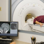 Read also: What does MRI of the brain show
Open article

The list was formed on the basis of the lack of convincing data on the effectiveness of drugs for the stated indications, as required by evidence-based medicine, as well as the absence of authoritative sources and recommendations.