What does a brain MRI show?
In the twentieth century, when high-level medical technologies were just entering society, identifying brain diseases was extremely difficult. An MRI machine came to the rescue, with which one can not only detect hidden pathologies, but also detect malignant neoplasms in the cerebral cortex.
Magnetic resonance imaging procedure and influencing factor
Before the study, it is important to understand the significance of the process and the ability to detect a brain tumor at an early stage . The diagnosis involves the physical phenomenon of magnetic resonance, which can somewhat affect the general well-being of the patient and his psyche, since maximum immobility is required when taking photographs.
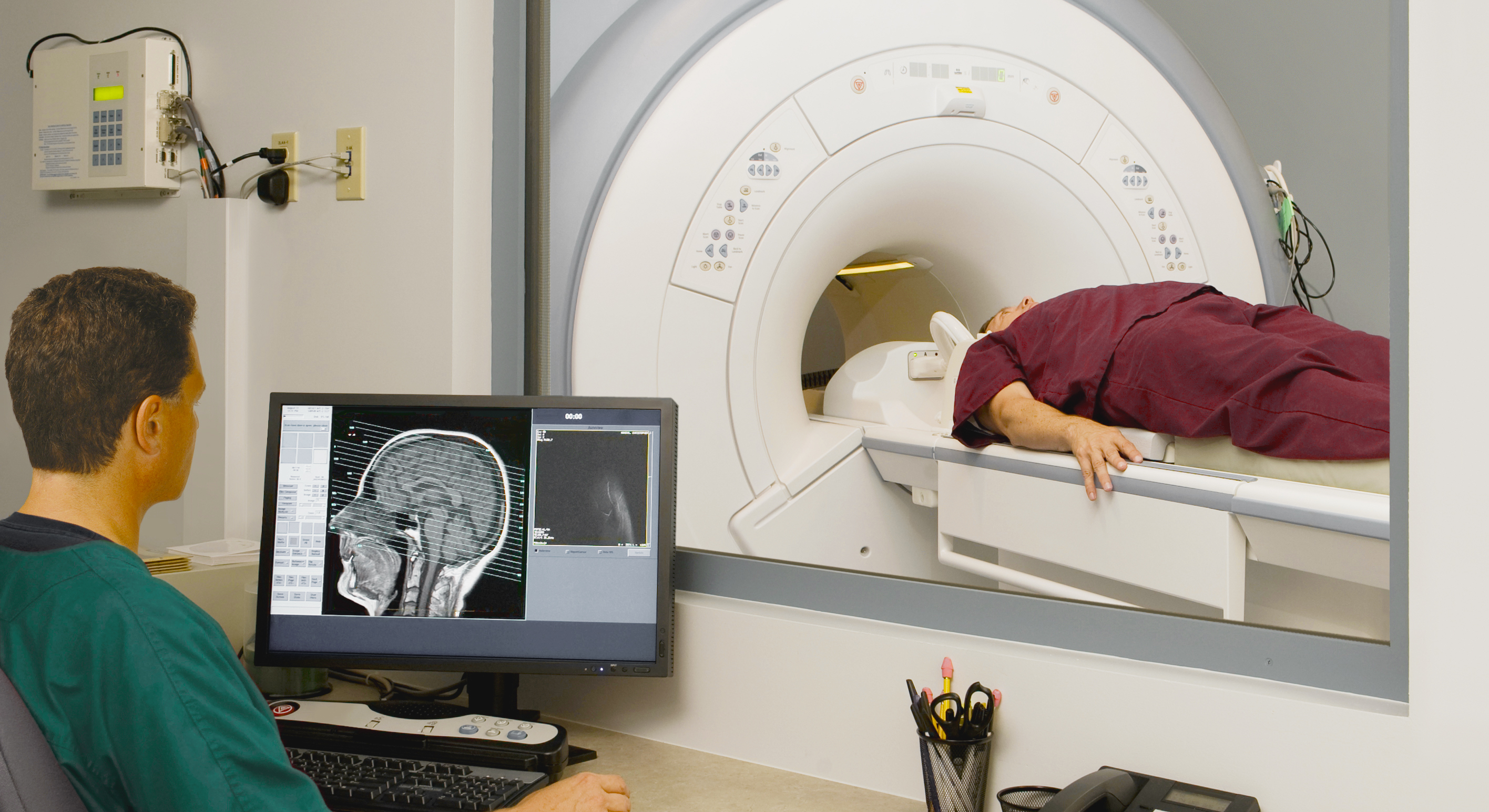
Such an examination has practically no contraindications, but MRI testing for negative effects in certain diseases is still ongoing, so you need to notify your doctor in advance about the characteristics of the body, including if you have recently had surgery using implants. The patient is placed on a gurney inside the device in a closed space; an undesirable effect may occur if there are mental deviations and phobias. In such cases, a sedative is prescribed.
The operation of the device causes noticeable noise and requires the maximum possible immobility of the area under study, for which it is secured with belts. The procedure can take quite a long time. For example, when taking readings from the head, it takes from 20 to 50 minutes, depending on the type of study. After this, the resulting images are analyzed by a radiologist, who makes a conclusion and refers him to a doctor.
Main indications for use
CT have no equal in detecting defects in brain function Obtaining accurate initial data when using this method is a qualitative characteristic of the procedure. An MRI of the head may be needed if it is necessary to detect:
- Deviations in the functioning of cerebral vessels
- Possible consequences of injuries and bruises
- Neoplasms or brain tumor
- Processes that cause regular pain attacks
- Problems with the speech apparatus and hearing defects
- Infectious infections and inflammatory processes
- Chronic diseases of the nose and middle ear
- Disorders and disorders of the nervous system, epilepsy
- Increased pressure inside the skull
- Anomalies of the development of the cranium
To obtain a brighter and most accurate picture, a contrast method can be used, while special drugs are introduced into the patient’s body, which can sometimes cause allergies.
A competent consultation with a specialist eliminates the occurrence of undesirable circumstances after an MRI diagnosis, and the patient’s openness in this case is absolutely necessary.
When examining the results of an MRI of the brain, the doctor receives a complete picture of the situation with an image of all parts of the brain that play an important role in the life of the brain. Congenital pathologies and hidden tumors will not escape an experienced eye, therefore, in modern diagnostic methods, MRI occupies a special niche and is an indispensable assistant in the process of treating the most important component of the human body.
Please rate the article:
 (2 ratings, average: 5,00 out of 5)
-
 Read also: 9 myths about brain tumors that you don’t need to believe
Open article

The list was formed on the basis of the lack of convincing data on the effectiveness of drugs for the stated indications, as required by evidence-based medicine, as well as the absence of authoritative sources and recommendations.


