Vascular brain tumor
The content of the article
Conventionally, such formations can be classified into cavernous and venous tumors. Multiple locations of angiomas are already angiomatosis. Angioma represents the greatest danger among formations. Cavernous angioma is a ticking time bomb. A dangerous complication of this disease is the possibility of rupture of the caverns with subsequent hemorrhage.
Causes
The reasons for the occurrence can only be speculative. Among them, genetic abnormalities, post-traumatic complications, and viral lesions of the lining of the brain dominate. Statistics show that concomitant pathologies, for example, cirrhosis of the liver, can also provoke the appearance of angiomas. The pathogenesis of angiomas is due to the tendency to dilate blood vessels and enlarge the lymph nodes. Young patients (children) are most prone to such diseases due to the failure of internal organs and the pathology of the development of certain systems. The risk of brain stem disease is significantly increased by various strokes.
Symptoms
Angioma can last for some time without noticeable signs.
They are often duplicated with symptom complexes of minor neurological abnormalities. The manifestation and intensity depends primarily on the location and size of the neoplasms. The most common symptoms: • Increasing or bursting headache ;
• Ringing and noise in the ears;
• Development of seizures;
• Paresis;
• Paralysis;
• Flattening of sensations;
• Speech disorder;
• Noticeable torpidity of thinking;
• Visual defects;
• Convulsions.
Diagnostics
Considering the fact that in 78% of cases the disease is asymptomatic, the process of diagnostic examination is very difficult.
Medical tactics work successfully only when the tumor, by its size and location, injures the blood vessels of the brain, depriving vital organs of normal blood supply. Nowadays, modern diagnostic methods are able to detect the disease at the initial stage:
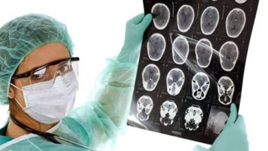
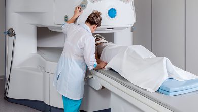
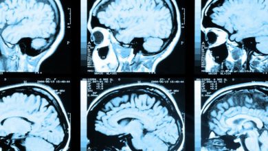
- Angiography;
- CT;
- MRI is a more detailed examination, based on magnetic and radio waves. The diagnostic method is more informative, with the least harm to health (X-rays are not used).
Treatment
Based on modern instrumental diagnostic data, taking into account the medical history, duration and clinical picture of the disease, the neurosurgeon selects the optimal treatment method:
- Surgically. A common method of getting rid of unwanted formations. Requires blockade of the blood supply before resection. It is possible to remove formations located relatively close to the surface.
- If the tumor is deep, it is appropriate to use the low-traumatic radio wave method. In this case, the blood vessels are soldered and the blood supply to the tumor tissue is stopped. Thus, its growth stops and the danger of hemorrhage is neutralized.
- Introduction of a sclerosing agent through a catheter. The blood vessels are clogged and tumor tissues are neutralized.
Unlike cavernous tumors, venous tumors of the cerebral vessels have a significantly better prognosis. In order to reduce undesirable deviations in the neurological status, it is necessary to carefully monitor the size and location of the tumor and carry out treatment at earlier stages. For preventive purposes, you need to undergo medical examinations as planned, as well as receive infusion treatment to strengthen the walls of blood vessels.
Please rate the article:
 (2 ratings, average: 5,00 out of 5)
-
 Read also: Chordoma - description and symptoms of the disease, prevention and treatment
Open article

The list was formed on the basis of the lack of convincing data on the effectiveness of drugs for the stated indications, as required by evidence-based medicine, as well as the absence of authoritative sources and recommendations.