Chordoma - description and symptoms of the disease, prevention and treatment
The content of the article
What is Chordoma?
Chordoma is a rare tumor, the source of which is the embryonic remnants of the notochord. The probability of its occurrence is considered to be 1-3 cases of the disease per million people per year. There is still no clear opinion among doctors and scientists regarding the classification.
The tumor rarely metastasizes and is characterized by slow growth, so it is often classified as benign. At the same time, cerebral chordoma is rarely accessible for effective treatment, provoking the development of serious complications and multiple relapses, which is why in most sources it is called a malignant tumor.
Causes of the disease
Pathology is diagnosed at any age, more often in the male population. It should be noted that chordomas in the brain region are detected more at a young age, sacral ones - in older people.
The specific causes of the neoplasm are not yet known, and the risk groups have not been described. Since cases of tumor are not often observed when a patient has a positive diagnosis from his relatives, the hereditary nature of the disease has not been proven.
Symptoms of Chordoma
The clinical picture is determined by the location of the pathological focus. Its cranial location can lead to further spread of the process into the area of the sella turcica, orbit, and nasopharynx. Damage to the hypoglossal, vagus and glossopharyngeal nerves leads to bulbar disorders. The main features include:
- regular headaches;
- swallowing problems that last for years;
- changes in vision that develop over a long period of time;
- deterioration of speech function.
Diagnosis of Chordoma
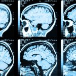
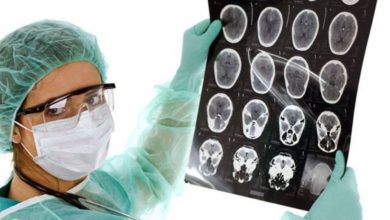
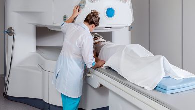
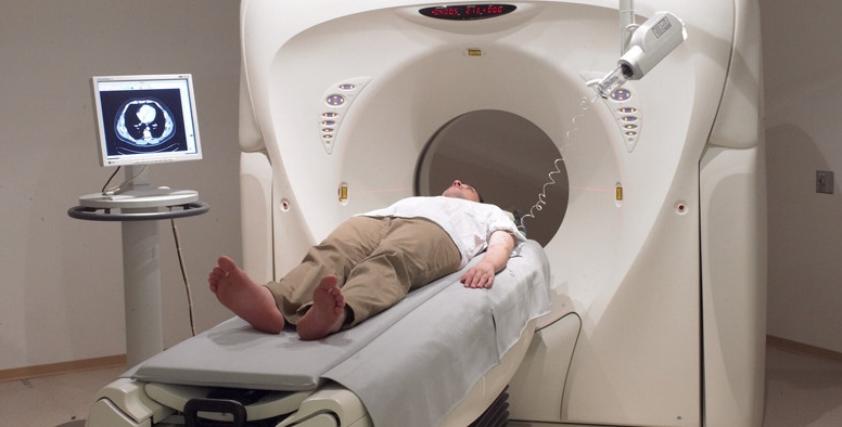
Important diagnostic procedures include neurological techniques and imaging methods, such as MRI , radiography, CT. The neurologist assesses the state of reflexes, motor activity in the limbs, and skin sensitivity to identify the focal clinical picture and preliminary establish the location of the pathological focus. During early diagnosis, a standard plain radiograph is taken, which makes it possible to identify destructive foci of bone tissue with a high degree of probability. According to statistics, in approximately half of the cases, during a standard x-ray examination, the doctor is able to notice calcifications inside the lesion.
To clarify the diagnosis, additional methods are prescribed - PET and scintigraphy . Among modern techniques that help determine the degree of damage to the body by chordoma, pneumoencephalography and angiography should be mentioned. Reliable methods include biopsy performed under the guidance of computed tomography or x-ray.
Treatment of Chordoma
When a diagnosis of cerebral chordoma is made, the prognosis remains unfavorable, even with the use of surgical treatment methods. The reason for this is the frequent recurrence of the process. Surgical intervention is made difficult by the proximity of the lesion to vital structures. In modern surgical practice, complete excision of the tumor simultaneously with the capsule is accepted. If this is not done, the likelihood of metastasis and relapse increases. Long-term medical practice confirms the formation of long-term remissions when the lesion was incompletely removed.
The first method is not highly effective and is used during an aggressive process. Some scientific sources note the radioresistance of the tumors in question. At the same time, modern treatment methods lead to significant positive changes in the course of the disease.
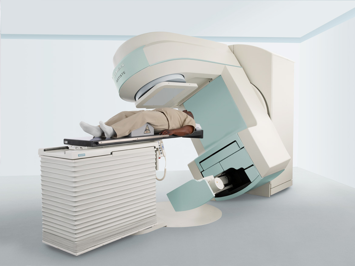
Radiation therapy
Radiation therapy can be prescribed in isolation and in combination with surgical techniques before and after their use. The preoperative procedure is done to reduce the size of the pathological lesion, so that it is then more convenient to remove the tumor, the postoperative procedure is done to completely destroy cancer cells in order to minimize relapses. After subtotal removal of a well-differentiated chordoma, in some cases, radiotherapy is not prescribed, but the observation process continues with periodic re-examination for six months using CT or MRI . For moderately or poorly differentiated chordoma, stereotactic radiotherapy is prescribed soon after surgery for subtotal and partial removal.
Radiation therapy is prescribed to patients when metastases are detected and if it is impossible for various reasons (severe general condition, inaccessible location of the lesion, high risk of complications) to carry out surgical treatment. Conventionally, the disease is called “radioresistant” because the tumor is not sensitive to radiation therapy in medium doses. The problem here is the delivery of significant doses of energy to relatively large volumes of the pathological focus with high accuracy.
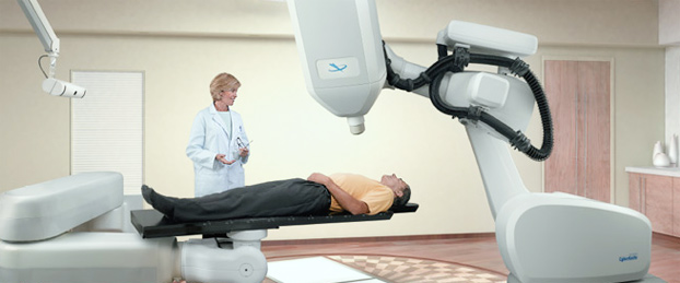
Cyber Knife system
In modern conditions, stereotactic radiation therapy is increasingly being practiced for the effective treatment of tumors of the central nervous system , as is radiosurgery. Modern techniques have already been successfully tested in the Gamma Knife and Cyber Knife systems, on specialized linear accelerators in three-dimensional planning and the use of multileaf collimators.
The general meaning of stereotactic radiation therapy is the highly targeted action of radiation beams on the tumor process from different directions. In this case, a high dose of radiation is formed directly at the epicenter of the outbreak, while healthy tissues around experience the most minimal load, which does not cause damage.
There is also no problem of patient displacement during treatment, which was effectively solved by the developers of the CyberKnife system. Human movements, including breathing, do not interfere with the system’s delivery of radiation beams to the target with extreme precision. In this case, the target of influence is under systematic control throughout the entire treatment period. As evidenced by numerous experimental data, the accuracy of the Cyber Knife reaches 0.5 mm.
There is only one limitation to the use of the CyberKnife system - the dimensions of the primary or residual lesion with a diameter of less than 3.5 cm. The effective technique is performed without special preparation of the patient, who after irradiation can immediately leave the clinic, receiving another 1-2 weeks of drug therapy. The clinical result after the technique is most often observed within several weeks. Partial regression of the tumor is observed within several months, and after a year and a half, the tumor process may disappear completely. Structural residual changes will be manifested by the absence of contrast accumulation.
Video of the operation (in English)
Patient observation
If necessary, the doctor can re-prescribe a course of radiotherapy using the CyberKnife system. Patients after subtotal removal of a well-differentiated tumor are subsequently shown dynamic observation; after six months, a repeat MRI or CT scan is performed. stereotactic is performed after surgery . If the chordoma was not completely removed, a consultation with a radiologist is required no later than three months after surgery.
Please rate the article:
 (3 ratings, average: 5,00 out of 5)
-
 Read also: Symptoms of a brain tumor
Open article

The list was formed on the basis of the lack of convincing data on the effectiveness of drugs for the stated indications, as required by evidence-based medicine, as well as the absence of authoritative sources and recommendations.

