Brain glioma, what is it?
The content of the article
Quite often, unfortunately, this disease occurs in people of all ages, so it is necessary to clearly understand it.
It varies in its degree of malignancy, age of manifestation, ability to invade and progress, and histological features.
Types of Gliomas
Gliomas are of the following types:
- Astrocytoma, an astrocytic glioma, is the most common and forms in the white matter. There are fibrillar, anaplastic astrocytomas , glioblastomas, pilocytic astrocytomas, pleomorphic .
- Oligodendroglioma arises from oligodendrocytes and occurs in 10% of diseases. These include oligodendrogliomas and anaplastic oligodendrogliomas.
- Ependymoma is formed in the ventricular system of the brain and occurs in 5-8% of brain diseases.
- Neuroma occurs in 8-9% of cancer cases.
- Mixed gliomas. These include oligoastrocytomas and anaplastic oligoastrocytomas.
- Tumors of the choroid plexus occur very rarely, accounting for 1-2% of the total number of cancers.
- Gliomatosis of the brain.
- Neuroepithelial tumors of unknown origin. These include astroblastomas and polar spongioblastomas.
- Neuronal and mixed neuronal-glial tumors are a very rare type of glioma, found in 0.5% of cases of cancer, these include gangliocytomas, gangliogliomas, neurocytomas, neuroblastomas, neuroepitheliomas.
There are several assumptions about what causes a glial brain tumor. The first is that astrocytomas develop from an astrocytic lineage, and oligodendrogliomas from an oligodendroglial lineage. Second, due to the presence of zones of malignant vulnerability, gliomas develop from slowly proliferating cells in which malignant transformation occurs, and not from mature cells. And the type of disease that occurs depends on the failure of certain types of genes.
There is a WHO classification of 4 degrees of malignancy of brain gliomas:
- Grade 1 - benign, grows slowly, the patient has a long life expectancy.
- Grade 2 - borderline gliomas, grow slowly, can progress to grade 3, 4.
- Grade 3 - malignant gliomas.
- Grade 4 - fast-growing malignant gliomas, the patient's life expectancy is short.
Glial brain tumor, symptoms
- Headache , which does not go away after taking painkillers, is accompanied by a feeling of heaviness in the eyes, nausea, vomiting, and sometimes even convulsive seizures. These signs are characteristic of diffuse glioma of the frontal part of the organ.
- Visual impairment.
- Violation of the vestibular system (dizziness, staggering when walking)
- Speech impairment.
- Impaired sensitivity.
- Mental abnormalities (impaired behavior, thinking, memory).
- Diffuse glioma of the pons and medulla oblongata manifests itself in the form of mental disorders, high intracranial pressure, vomiting, paralysis of the vocal cords, and with malignant glioma of the right half of the medulla oblongata, auditory, visual and taste hallucinations appear.
- With diffuse glioma of the midbrain tegmentum, fine motor skills are impaired, and sometimes paralysis of the limbs occurs.
- With diffuse glioma of the midbrain, disturbances in the motor functions of the body appear, orientation in space is lost, and the ability to move without the help of others is lost.
- Disease of the corpus callosum manifests itself in impaired coordination of movements, mental disorders, and loss of the ability to write and draw.
- Cramps.
- Weakness in arms and legs.
- Personality change.
- Diffuse glioma of the brainstem in children, the prognosis for which is disappointing, with a high mortality rate. This is a widespread neoplasm in this part of the head and is very dangerous.
There is also a microscopic variety of gliomas. When performing tissue examination, gliomas are divided into:
- Protoplasmic. Occurs in the cells of the gray matter, and may degenerate into a cystic form. This is a benign type of tumor, characterized by local growth.
- Fibrillar occurs in the white matter and also has many massive nodes prone to necrosis. It is also a benign tumor.
- Anaplastic is prone to transition to malignant, characterized by the presence of multinucleated cells with pathological changes and deformation of the nuclei.
This disease is a spherical or oval elongated structure with blurry indistinct boundaries, has a gray-pink or white color, sometimes red. The size of the tumor can range from a few millimeters to 10 centimeters in diameter.
How does brain glioma affect life expectancy?
Life expectancy with glioma largely depends on the rate of tumor growth and the intensity of clinical signs. The average lifespan is more than 5 years with timely diagnosis and proper treatment of the disease, as well as the general well-being of the person and his attitude to fight this disease. Of course, when a tumor is detected already at stage 4, treatment may even be completely useless. No matter how the patient struggles, life expectancy will be short.
Gliomas can be benign or malignant. But in any case, when a diagnosis of brain glioma is made, the life prognosis will not be positive, because whatever the degree of malignancy of the tumor, it still affects the normal functioning of the organ to a greater or lesser extent, which leads to various disturbances in the activity of the entire human body.
For a long time, the average life expectancy of patients with glioma was only about 12 months from the time of diagnosis. At the moment, thanks to the use of the latest technologies, drugs in combination with radiation therapy, the life expectancy of patients has begun to reach a five-year level.
Glioma grade 3
For stage 3 diseases, the prognosis, unfortunately, is not very favorable. Even surgery in most cases does not guarantee a cure. And life expectancy directly depends on the success of the operation and the number of lobes removed.
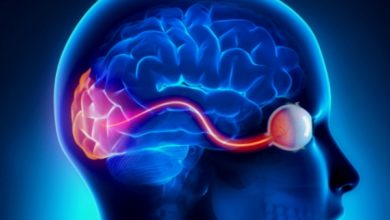
When glioma develops, symptoms at the very beginning of the disease can be almost invisible. Sometimes, there are no signs of the disease, and the tumor is discovered completely by accident. If a person suspects a brain tumor, they should immediately seek advice from a qualified specialist, talk about all the signs and symptoms and undergo a full medical examination. In addition to the general examination, it will be necessary to undergo an examination by a neurologist, during which the doctor will check how the patient speaks and moves. In addition, the specialist will need to conduct an examination of the patient's eyes to determine the location of the tumor and its possible effect on the optic nerve. The optic nerve connects the eyes to the brain, and when glioma develops, it becomes inflamed and swollen. If such signs are found, the patient will need immediate medical attention.
Diagnostics
Diagnosis of glioma consists of several stages:
- medical examination, examination of anamnesis, during which a specialist asks about warning symptoms and clarifies what diseases the patient or his relatives have suffered;
- examination by a neurologist, which can help clarify some problems: with vision, hearing, coordination of movements, balance, reflexes, thinking and memory;
- examination of the brain using magnetic resonance imaging (MRI) or computed tomography (CT), which is the most common method for diagnosing this disease;
- A biopsy is a procedure in which small pieces of the tumor are removed to be examined in more detail and analyzed under a microscope.
Low-grade glioma, what is it?
Low-grade glioma (LGG) is a neoplasm, grade 1-2 tumor according to the WHO classification.
There are several groups of low-grade gliomas that differ by cell lineage:
- Infiltrative astrocytoma grade 2 (fibrillar and protoplasmic).
- Oligodendroglia from astrocytes and oligodendroglia (oligoastrocytomas).
This disease is divided into 3 groups, regardless of histology:
- Solid tumors that do not infiltrate the brain tissue. Most often, when such a neoplasm forms, surgical intervention is possible, removal occurs completely, and the prognosis for life expectancy is most favorable. These include gangliogliomas, pilocytic astrocytomas, pleomorphic xanthoastrocytomas, and some protoplasmic astrocytomas (oligodendrogliomas do not belong to this group).
- Dense formations that are surrounded by a zone of infiltration of brain tissue. Surgical intervention is possible, but complete removal can only be done from the site of origin. They exist as low-grade astrocytomas.
- Infiltration by tumor cells, no tumor node. Due to the risk of neurological deficits, resection of infected tissue may not be possible. Forms as an oligodendroglioma .
Diffuse glioma of the brainstem in children
The brain stem is the connection between the spinal cord and the brain, located at the end of the spinal cord. Responsible for the functioning of the most important systems in the human body - cardiac, motor and respiratory. Also responsible for thermoregulation of the body and metabolism.
of the brain stem responsible for these functions : the middle, oblongata, intermediate and pons. A tumor can arise in any of these departments and the impact on various functions in the patient’s body depends on the location of its appearance. In most cases, it occurs on the pons, a tumor of the medulla oblongata is less common, and is least common in the midbrain.
Brain stem glioma in children, first symptoms
Symptoms may not appear for a long time, but from the moment they arise they intensify quite quickly. In most cases, they are added gradually and therefore the disease does not manifest itself for a long time.
Main symptoms:
- impaired breathing and functioning of the cardiovascular system (myasthenic syndrome, respiratory failure, tachyapnea, bradycardia). Occurs in almost half of tumor cases and can lead to death.
- nystagmus (eye twitching), facial asymmetry due to weakness of facial muscles. Occurs in 50% of cases.
- problems with vision and hearing (double vision, paresis of the eye muscles and deafness, strabismus, tinnitus) occur in 20% of patients.
- dysphagia and speech impairment, occurs in 15% of patients, occurs due to paresis of the muscles of the tongue, larynx, and palate. As a result, it is painful for the child to swallow food and it is difficult to talk, the voice becomes muffled, and words are blurred.
- trembling in the hands, decreased overall muscle tone - occurs in 40% of patients.
- impaired coordination of movements, unsteady gait, weakness in the lower extremities.
There are also general symptoms of the disease:
- Fatigue, drowsiness, absent-mindedness, memory impairment.
- Changes in behavior, aloofness, irritability, withdrawal.
Hydrocephalus (fluid accumulation in the brain) appears in the final stages, and sometimes does not appear at all. Expressed by the following symptoms:
- headache that gets worse when you move your head, cough or sneeze;
- nausea, vomiting;
- dizziness.
Treatment of grade 1-2 gliomas
When treating grade 1 and 2 benign glioma, a complex of measures is used, which includes surgery, chemotherapy and radiation. Surgery alone does not help completely get rid of the tumor. To completely destroy it, radiation therapy is used.
Endoscopy is also used. In this case, the infected tissue is removed using an endoscope, which is inserted into the skull along with surgical instruments and a video camera, with which you can monitor the process of surgical manipulations. During surgery, a larger amount of the tumor is excised, which eliminates compression of the brain tissue, restores liver circulation, stabilizes intracranial pressure, reduces swelling of the organ, and improves the patient’s condition. After this, a course of chemotherapy is carried out. Surgery is also explained by the fact that the inner layers of the tumor are not susceptible to the influence of drugs and even ionizing radiation. After this, constant monitoring of the patient’s condition is required; complications may arise, which manifest themselves in the form of swelling of the soft tissues of the eyelids and forehead, intracranial hemorrhage, infection of the skull tissue, which, in turn, leads to the development of meningitis and encephalitis.
Treatment of a benign tumor
The only difference between the treatment of a benign tumor and a malignant one is that the former does not require chemotherapy. The general treatment plan is developed by the doctor personally for each patient, depending on the age, location of the tumor and the general condition of the patient.
The main principle of treatment for benign glioma is craniotomy - the skull is opened and the tumor is excised, and then doctors administer a course of radiation therapy. In most cases, it is carried out in the traditional way - remotely or in the form of proton therapy or radio surgery (using a gamma knife and a cyber knife).
Currently, the latest technologies are being introduced to combat this type of brain cancer. The robotic cyber-knife system is becoming widespread. It compares favorably with others in a number of advantages, for example, the absence of harmful effects on the entire patient’s body and the ability to remove tumors even in the most inaccessible places.
Corticosteroids are used for drug treatment of brain tumors because they can reduce swelling of brain tissue.
Incurable benign gliomas are very rare.
In almost 70% of cases, patients experience an improvement in their condition after surgery. Although there are cases of some consequences that manifest themselves in the form of decreased vision, a drop in general tone and difficulty speaking. After operations to remove gliomas, 50% of adult patients aged 20 to 44 years had normal five-year survival rates, and in those over 65 years of age this figure was reduced to 5%.
Here's what you need to know about cancer in the form of brain glioma, monitor your health and, if you have suspicious symptoms, play it safe and immediately seek advice from specialists. Be less nervous, overtired, sleep more and enjoy life!
Please rate the article:

 (4 ratings, average: 4,75 out of 5)
-
 Read also: Chiasmal glioma - symptoms, diagnosis and treatment
Open article

The list was formed on the basis of the lack of convincing data on the effectiveness of drugs for the stated indications, as required by evidence-based medicine, as well as the absence of authoritative sources and recommendations.